This comprehensive article explores the intricate neurological connections between vertigo, vomiting, and nausea. We’ll examine how these seemingly distinct symptoms share complex neurological pathways, their various causes ranging from peripheral inner ear disorders to central nervous system conditions, and the latest approaches to diagnosis and treatment. Understanding these connections can lead to more effective management strategies and improved quality of life for affected patients.
The Neurological Basis of Vertigo
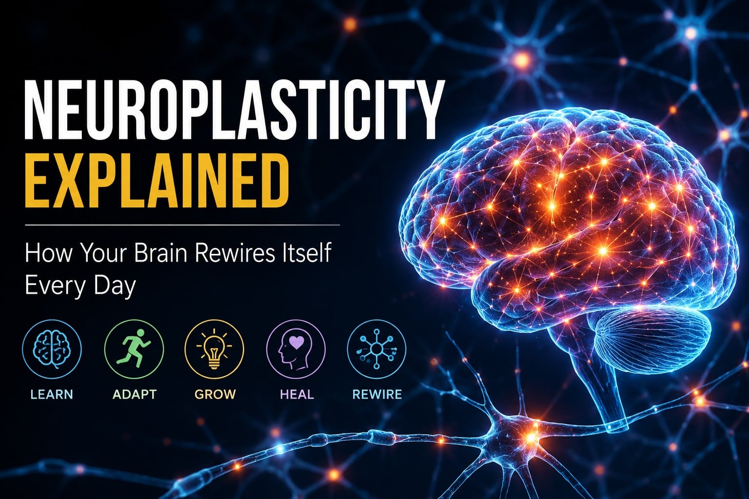
Vertigo is not merely dizziness but a specific sensation of spinning or movement when none is occurring. This disorienting symptom stems from sophisticated neural mechanisms centered on the vestibular system, which plays a crucial role in maintaining balance and spatial orientation. Located within the inner ear, the vestibular apparatus contains three semicircular canals filled with fluid and tiny hair cells that detect rotational movements, plus the utricle and saccule that sense linear accelerations and head position relative to gravity.
The eighth cranial nerve (vestibulocochlear nerve) serves as the primary communication channel between the vestibular apparatus and the brain. It transmits signals from the inner ear to the vestibular nuclei in the brainstem, where initial processing occurs. From there, information travels to multiple brain regions including the cerebellum, thalamus, and cerebral cortex. This complex network allows for the integration of vestibular input with visual and proprioceptive signals to maintain spatial awareness and equilibrium.
Key Components of Balance
- Vestibular system (inner ear)
- Visual system
- Proprioception (position sense)
- Cerebellar coordination
Neural Pathways
- Vestibular nuclei in brainstem
- Vestibulocerebellar tracts
- Vestibulospinal tracts
- Vestibulo-ocular connections
When discrepancies arise between these sensory inputs or when the vestibular system itself malfunctions, the brain receives conflicting information about the body’s position and movement. This sensory mismatch is interpreted as vertigo. The intense disorientation from vertigo often activates additional neurological pathways connected to autonomic responses, which explains why vertigo rarely occurs in isolation and frequently triggers accompanying symptoms like nausea and vomiting.
How the Brain Links Vertigo to Nausea and Vomiting
The neurological connection between vertigo, nausea, and vomiting represents one of the most fascinating examples of interconnected neural pathways in human physiology. When the vestibular system sends abnormal or conflicting signals to the brain—as occurs during vertigo—these signals don’t remain isolated within balance-processing circuits. Instead, they activate the brain’s protective mechanisms against potential toxins or spatial disorientation.
- Sensory Conflict Detection: Brain detects mismatch between vestibular, visual, and proprioceptive inputs
- Neural Pathway Activation: Signals travel to vestibular nuclei and on to the chemoreceptor trigger zone (CTZ)
- Autonomic Response: Activation of vomiting center in medulla initiates nausea and emetic reflex
- Feedback Loop: Continued vestibular disturbance maintains the cycle of symptoms
The area postrema, or chemoreceptor trigger zone (CTZ), located in the medulla oblongata, plays a pivotal role in this connection. While primarily sensitive to toxins in the bloodstream, it also receives input from the vestibular system. When the CTZ is activated by abnormal vestibular signals, it stimulates the nearby vomiting center, also in the medulla. This vomiting center coordinates the complex sequence of muscular contractions involved in the emetic response.
From an evolutionary perspective, this connection makes sense. The same nausea and vomiting that protects us from ingested toxins also activates during spatial disorientation, which historically might indicate exposure to neurotoxins or dangerous movement situations. Essentially, the brain interprets conflicting vestibular information as a potential threat and initiates protective responses accordingly. This explains why medications that block certain neurotransmitters (particularly histamine and acetylcholine) can effectively treat both vertigo and its associated nausea, as they interrupt these shared neurological pathways.
Peripheral Causes: Inner Ear Disorders
Peripheral vertigo arises from dysfunction within the inner ear’s vestibular apparatus rather than in the central nervous system. These disorders account for approximately 80% of all vertigo cases and typically produce intense but self-limited episodes. Understanding their distinct mechanisms helps guide appropriate treatment approaches.

Benign Paroxysmal Positional Vertigo (BPPV)
BPPV represents the most common cause of peripheral vertigo, affecting up to 2.4% of the population at some point in their lives. This condition occurs when calcium carbonate crystals (otoconia) dislodge from their normal location in the utricle and migrate into the semicircular canals, most commonly the posterior canal. These misplaced crystals disrupt the normal fluid dynamics in the canals, causing inappropriate triggering of vestibular hair cells with certain head movements. The resulting neural signals create a false sensation of rotation, typically lasting seconds to minutes. BPPV classically produces brief, severe vertigo triggered by specific head positions, such as rolling over in bed or looking up.
Vestibular Neuritis and Labyrinthitis
These inflammatory conditions affect either the vestibular nerve (neuritis) or both the nerve and labyrinth (labyrinthitis). Typically triggered by viral infections—often the same viruses responsible for upper respiratory infections—these conditions cause sudden-onset, severe vertigo lasting days to weeks. The inflammation disrupts normal signal transmission from the affected ear to the brain, creating a significant vestibular asymmetry that the brain interprets as constant rotation. Unlike BPPV, these conditions often present with constant vertigo regardless of position, severe nausea and vomiting, and possible hearing loss (in labyrinthitis).
Meniere’s Disease
This chronic condition involves endolymphatic hydrops—an excessive accumulation of endolymph fluid in the inner ear. The exact cause remains unclear, but the increased pressure disrupts normal vestibular and cochlear function. Meniere’s disease typically presents with recurrent episodes of severe vertigo lasting hours, fluctuating hearing loss, tinnitus, and aural fullness. The unpredictable nature of attacks and the constellation of symptoms make this condition particularly challenging for patients.
Central Neurological Causes
Central causes of vertigo originate within the brain itself rather than the peripheral vestibular system. These conditions typically involve the vestibular nuclei in the brainstem, the cerebellum, or higher cortical areas that process balance information. While less common than peripheral causes, central vertigo often indicates more serious underlying neurological conditions requiring prompt medical attention.
Vestibular Migraine
The most common central cause of recurrent vertigo, affecting approximately 1% of the general population and up to 10% of migraine sufferers. Unlike traditional migraines, vestibular migraines may present without headache, making diagnosis challenging. The pathophysiology involves abnormal neuronal excitability, cortical spreading depression, and trigeminal-vestibular interactions. During attacks, abnormal processing of vestibular information in the brainstem and cortical regions creates vertigo episodes lasting minutes to days.
Cerebellar Disorders
The cerebellum plays a crucial role in motor coordination and vestibular processing. Lesions affecting the cerebellum often produce persistent dizziness, gait ataxia, and nystagmus with specific directional characteristics. Common causes include strokes, multiple sclerosis plaques, tumors, or degenerative cerebellar diseases. These conditions typically present with additional neurological symptoms beyond vertigo itself, such as dysarthria, limb incoordination, or diplopia.
Brainstem Disorders
The vestibular nuclei in the brainstem serve as the primary relay stations for balance information. Lesions here, particularly from ischemic strokes or demyelinating diseases, can produce severe vertigo accompanied by crossed neurological findings (e.g., facial numbness on one side with limb weakness on the opposite side). These conditions warrant immediate medical evaluation as they may represent acute stroke or other life-threatening conditions.
Central vertigo often differs from peripheral vertigo in several key ways. The onset may be more gradual, the intensity potentially less severe but more persistent, and the associated symptoms frequently include other neurological findings. Importantly, central vertigo is less likely to respond to typical vestibular suppressant medications. Neuroimaging, typically with MRI, plays a crucial role in diagnosing these conditions, allowing visualization of structural abnormalities in the brain that may be responsible for the symptoms.
The connection between central vertigo causes and accompanying nausea/vomiting relates to direct activation of the chemoreceptor trigger zone and vomiting center in the medulla. When pathology affects areas near these centers, the threshold for triggering the emetic response may be lowered, explaining why some central causes of vertigo produce particularly severe autonomic symptoms.
Symptom Profiles and Differential Diagnosis
Accurate diagnosis of vertigo requires careful analysis of symptom patterns and associated neurological findings. The distinction between peripheral and central causes is particularly crucial as it determines both the urgency of intervention and the appropriate treatment approach. Clinicians rely on specific symptom profiles and examination findings to guide this differential diagnosis.
| Feature | Peripheral Vertigo | Central Vertigo |
|---|---|---|
| Onset | Typically sudden | Often gradual |
| Intensity | Severe, often incapacitating | Usually less severe but more persistent |
| Duration | Seconds (BPPV) to hours (Meniere’s) | Often constant, may last days to weeks |
| Nystagmus | Horizontal-torsional, inhibited by visual fixation | Direction-changing, vertical, or pure torsional; not suppressed by fixation |
| Associated symptoms | Hearing loss, tinnitus, fullness in ear | Diplopia, dysarthria, dysphagia, limb ataxia, sensory disturbances |
| Nausea/vomiting | Common, often proportional to vertigo severity | Variable, may be disproportionate to vertigo |
Associated symptoms provide crucial diagnostic clues. Concurrent hearing loss, tinnitus, or aural fullness strongly suggests a peripheral cause (particularly Meniere’s disease or labyrinthitis). In contrast, accompanying diplopia, facial numbness, limb weakness, or speech difficulties points to central pathology requiring urgent evaluation.
67%
Present with Nausea
Percentage of vertigo patients who experience accompanying nausea
42%
Have Vomiting
Percentage of vertigo patients who progress to actual emesis
85%
Peripheral Cases
Approximate percentage of vertigo cases with peripheral origin
Several specific clinical tests help distinguish between different causes of vertigo. The Dix-Hallpike maneuver specifically tests for BPPV, producing characteristic delayed-onset, temporary nystagmus when the affected ear is positioned appropriately. The head impulse test assesses vestibulo-ocular reflex function, with abnormal catch-up saccades indicating peripheral vestibular dysfunction. The HINTS examination (Head Impulse, Nystagmus, Test of Skew) has proven particularly valuable in the acute setting, with sensitivity exceeding that of early MRI for stroke detection in acute vertigo.
Red flags warranting immediate neurological evaluation include vertical nystagmus, new-onset severe headache, any focal neurological deficits, or acute vertigo in patients with vascular risk factors. These findings raise concern for potentially serious central causes requiring prompt intervention, particularly vertebrobasilar stroke affecting the posterior circulation of the brain.
Management and Treatment Approaches
Treatment strategies for vertigo and its associated symptoms depend primarily on the underlying cause. Management approaches range from simple repositioning maneuvers for BPPV to medication regimens for symptomatic relief to comprehensive vestibular rehabilitation programs. Understanding the neurological basis of these symptoms guides the most effective therapeutic interventions.
- Accurate Diagnosis: Identifying the specific cause through history, examination, and testing
- Symptom Management: Medications and maneuvers to control acute symptoms.
- Vestibular Rehabilitation: Exercises to promote central compensation.
- Addressing Underlying Cause: Targeted treatment of specific neurological condition
Medication Approaches
Several medication classes can help manage vertigo and associated nausea/vomiting. Vestibular suppressants like meclizine, diazepam, or scopolamine reduce vestibular sensitivity and help control acute symptoms. Antiemetics such as promethazine, ondansetron, or metoclopramide target the chemoreceptor trigger zone and vomiting center directly to reduce nausea. Importantly, these medications should generally be used short-term for acute symptoms, as prolonged use can delay the brain’s natural compensation process. For specific conditions like vestibular migraine, preventive medications including topiramate, propranolol, or certain antidepressants may reduce attack frequency.
Repositioning Maneuvers
For BPPV, specific particle repositioning maneuvers—the Epley, Semont, or BBQ roll techniques—effectively relocate the displaced otoconia back to their proper location. These simple, non-invasive procedures have success rates exceeding 80% in properly diagnosed cases, often providing immediate relief. Some patients benefit from learning self-administered home versions of these maneuvers for recurrent episodes.
Vestibular Rehabilitation Therapy
This specialized form of physical therapy promotes central nervous system adaptation to vestibular dysfunction. Through carefully designed habituation, gaze stabilization, and balance training exercises, vestibular rehabilitation helps the brain recalibrate its interpretation of vestibular signals and compensate for deficits. This approach proves particularly valuable for patients with unilateral vestibular loss (e.g., after vestibular neuritis) or those with persistent symptoms despite appropriate treatment of the underlying cause. Evidence suggests that early initiation of vestibular rehabilitation leads to faster recovery and better long-term outcomes.
For cases with clear central neurological causes, treatment must target the underlying condition. This may involve antiepileptic medications for vestibular epilepsy, migraine prophylaxis for vestibular migraine, or specific interventions for structural lesions including surgical approaches when appropriate. The management of vertigo from central causes typically requires a multidisciplinary approach involving neurologists, otolaryngologists, and vestibular therapists working in concert.
Summary and Outlook
The intricate neurological connections between vertigo, nausea, and vomiting highlight the complex integration of vestibular, autonomic, and central nervous system functions. This document has explored how these seemingly distinct symptoms share common neural pathways, with vestibular disturbances frequently triggering activation of the brain’s emetic response mechanisms. We’ve examined how various conditions—from common benign peripheral disorders like BPPV to more concerning central pathologies like vertebrobasilar stroke—can disrupt these systems and produce characteristic symptom patterns.
The differential diagnosis between peripheral and central causes remains crucial for appropriate management. While peripheral vertigo typically presents with more severe but self-limited episodes often accompanied by auditory symptoms, central vertigo warrants particular vigilance due to its association with potentially serious neurological conditions. The presence of additional neurological symptoms, atypical nystagmus patterns, or red flag features should prompt immediate comprehensive evaluation.
Treatment approaches continue to evolve, with growing evidence supporting early vestibular rehabilitation for many conditions. Particle repositioning procedures remain remarkably effective for BPPV, while pharmacological interventions provide symptomatic relief for acute episodes across various conditions. For chronic or recurrent vertigo, addressing the underlying neurological cause becomes paramount.

Future Research Directions
- Advanced neuroimaging techniques to better visualize vestibular pathways
- Development of more selective antiemetics with fewer side effects
- Targeted biological therapies for immune-mediated vestibular disorders
- Virtual reality applications in vestibular rehabilitation
- Improved implantable devices for bilateral vestibular hypofunction
Early and accurate diagnosis, coupled with appropriate targeted treatment, remains the cornerstone of effective management for these neurologically interconnected symptoms.
The outlook for patients with vertigo continues to improve as our understanding of the underlying neurological mechanisms advances. Multidisciplinary approaches combining expertise from neurology, otolaryngology, and vestibular rehabilitation offer the most comprehensive management strategies. Patient education about the neurological basis of symptoms helps improve compliance with treatment regimens and reduces anxiety about potentially concerning symptoms. For most patients, particularly those with peripheral causes, the prognosis remains favorable with appropriate intervention, though some conditions like Meniere’s disease may require ongoing management strategies. As research continues to illuminate the complex neural connections between balance, nausea, and vomiting mechanisms, we can anticipate increasingly targeted and effective treatment approaches in the future.
At Hope Brain and Body in Chadds Ford, PA, we offer personalized treatment using Chiropractic Care alongside Wellness and Functional Neuro and now Stem Cell Therapy. To explore your options, Reach out to our team at Hope Brain & Body on (610) 652-4732 and schedule a visit to our offices in Chadds Ford, Pennsylvania. With ongoing advancements in understanding and managing adrenal POTS, patients can look forward to better symptom control and an improved overall well-being.