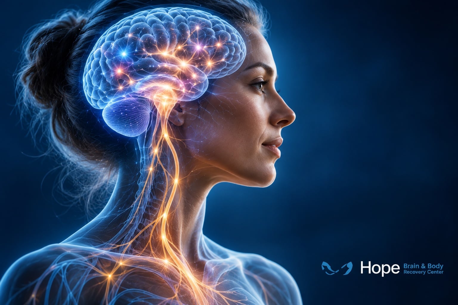
What if the brain could be encouraged to strengthen the exact neural connections it needs to recover?
Imagine someone recovering from a stroke who is relearning how to reach for a glass, open their hand, or button a shirt.
Every repetition sends signals through networks responsible for movement. Over time, practice may help those networks adapt.
But what if there were a way to enhance the brain’s response to that practice?
This is one of the most exciting questions being explored through vagus nerve stimulation (VNS).
A 2025 research paper by Kumagai, Shiramatsu, Kawai, and Takahashi, discussed in Frontiers in Neural Circuits, examines how VNS influences communication within brain circuits and why combining stimulation with specific activities may promote targeted neuroplasticity.
The central idea is fascinating: VNS may help reinforce the brain circuits that are active at the moment stimulation is delivered.
At Hope Brain & Body Recovery Center, understanding how the brain adapts to neurological stimulation is central to our approach to Brain-Specific Rehabilitation.
Let’s explore what happens inside the brain when the vagus nerve is stimulated—and why this research could matter for neurological recovery.
What Is Vagus Nerve Stimulation?
The vagus nerve is the tenth cranial nerve and an important communication pathway between the brainstem and internal organs.
It carries information involved in autonomic regulation, including aspects of heart function, digestion, and internal sensory processing.
But its connections also extend into brain networks involved in attention, learning, emotion, and movement.
Vagus nerve stimulation involves delivering controlled electrical impulses to influence activity along these pathways.
Some VNS treatments use surgically implanted devices, while research also explores noninvasive approaches that stimulate selected regions of the ear or neck.
Although VNS has established clinical applications in certain forms of epilepsy and treatment-resistant depression, researchers are increasingly interested in its potential to support neurological rehabilitation.
The important question isn’t simply whether stimulation activates the brain.
It’s how that activation changes the way neurons communicate.
Neuroplasticity: How the Brain Learns and Reorganizes
Neuroplasticity is the nervous system’s ability to change its structure and function in response to experience, activity, and injury.
Every time you learn a new skill, practice a movement, or adapt to a changing environment, neural networks participate in this process.
Some connections become more efficient.
Others become less influential.
Entire networks can change how they coordinate information.
This process is particularly important following neurological injury.
For example, when a stroke damages pathways involved in movement, rehabilitation aims to help the nervous system recover function through repeated, meaningful activity.
But not all stimulation produces useful plasticity.
The brain needs to reinforce the right connections at the right time.
That’s where VNS research becomes especially interesting.
The Core Discovery: Feedforward vs. Feedback Brain Signaling
The 2025 research presentation highlights a proposed mechanism involving the balance between feedforward and feedback processing.
These are two important ways that information moves through neural networks.
Feedforward Processing
Feedforward processing describes information moving through a network toward subsequent stages of processing.
For example, sensory information may travel from early sensory regions toward higher-order cortical areas.
Feedforward pathways help transmit incoming information and contribute to the brain’s response to events.
Feedback Processing
Feedback processing involves signals traveling from higher-order regions toward earlier processing areas.
These pathways help shape perception, expectations, attention, and the interpretation of incoming information.
Both systems are essential.
The research proposes that VNS can shift their relative influence, favoring feedforward signal transmission under certain conditions.
This offers a theoretical explanation for how stimulation may change information processing across interconnected brain circuits.
However, this is a proposed mechanistic model—not proof that every VNS protocol produces the same feedforward changes in every patient.
Two Chemical Messengers That Help the Brain Change
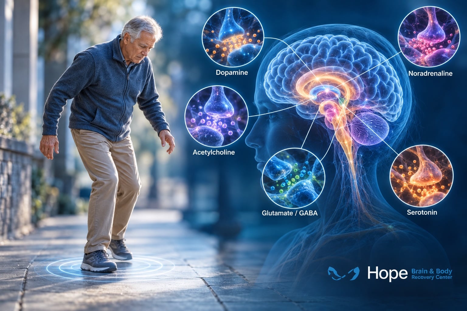
One of the most important aspects of VNS is its influence on neuromodulatory systems.
Neuromodulators are chemical messengers that influence how neurons respond to signals and how neural connections change.
The research focuses particularly on two systems:
Acetylcholine and norepinephrine (noradrenaline).
Both are involved in attention, learning, and neural plasticity.
Acetylcholine: Supporting Learning and Memory
Acetylcholine is associated with learning, memory, attention, and cortical plasticity.
An important source of acetylcholine in the brain is the basal forebrain cholinergic system.
The presentation explains that brief cholinergic activation can enhance plasticity when it coincides with relevant neural activity.
This means the timing of acetylcholine signaling may help determine which connections are strengthened.
Imagine practicing a difficult hand movement.
The motor networks involved in that movement become active.
If a neuromodulatory signal arrives at the right moment, it may help reinforce the activity occurring in those networks.
This is one reason researchers are interested in pairing VNS with rehabilitation rather than delivering stimulation independently of a task.
Norepinephrine: Helping the Brain Recognize Important Signals
The second major system involves norepinephrine.
Norepinephrine contributes to alertness, attention, learning, and the brain’s response to meaningful events.
A major source of norepinephrine is the locus coeruleus, a small structure located in the brainstem.
VNS can influence activity within this system.
The resulting norepinephrine signaling may help create conditions that support changes in neural connections.
Together, acetylcholine and norepinephrine provide a potential explanation for how VNS can influence learning across multiple brain networks.
The Locus Coeruleus: A Small Structure With a Major Role
The locus coeruleus is tiny compared with the cerebral cortex.
But its influence is widespread.
Its projections reach numerous brain regions involved in attention, memory, sensory processing, and motor control.
The research presentation describes experimental findings in which brief VNS trains lasting approximately 0.5 seconds produced rapid, phasic activation of locus coeruleus neurons.
Phasic activation refers to a short burst of activity rather than a continuously elevated signal.
In the experiments summarized, stimulation intensity and pulse width influenced the magnitude of the response.
These findings are important because they suggest that VNS can deliver precisely timed neuromodulatory signals.
That precision may be particularly useful when stimulation is paired with a movement or learning event.
However, these experimental parameters are not instructions for home stimulation. Clinical settings must be selected according to the device, indication, and patient’s medical circumstances.
Why Timing Matters More Than Simply Stimulating the Brain
This may be the most important concept in the entire research presentation.
Vagus nerve stimulation can have widespread neurological effects, but its influence on learning may become more specific when paired with a particular behavior.
Consider two scenarios.
In the first, stimulation is delivered without any particular movement or learning task.
In the second, stimulation is delivered repeatedly while someone practices a specific movement.
The second approach may help reinforce the neural circuits actively participating in that task.
The presentation describes this as event-specific plasticity.
Rather than strengthening every connection indiscriminately, paired stimulation may enhance the brain’s natural learning signals during meaningful behavior.
This helps explain why researchers are investigating VNS as an addition to structured neurological rehabilitation.
The stimulation doesn’t replace the exercise.
It may help the brain respond more effectively to the exercise.
How VNS May Improve Motor Learning
Motor learning involves more than muscle strength.
It requires the brain to coordinate sensory feedback, movement planning, timing, muscle activation, and the evaluation of movement outcomes.
When learning a new skill, the brain repeatedly compares intended movement with actual performance.
Successful movement patterns can gradually become more efficient.
The research presentation describes experimental studies in which VNS influenced activity within the primary motor cortex, a region involved in generating voluntary movements.
When stimulation was paired with skilled motor behavior, it altered the timing of neural responses associated with behavioral outcomes.
These changes were linked to the consolidation of learned movement patterns.
The proposed mechanism involves cholinergic reinforcement of circuits engaged during the task.
This is particularly relevant for rehabilitation because functional recovery often depends on relearning specific movements—not simply increasing overall brain activity.
Why VNS-Paired Rehabilitation Is Different From Ordinary Stimulation
Imagine a patient working with a therapist to regain hand control after a stroke.
The patient repeatedly practices opening the hand, grasping an object, and releasing it.
Each movement activates specific sensorimotor pathways.
In a VNS-paired rehabilitation protocol, stimulation is coordinated with selected repetitions.
The objective is to reinforce neural activity associated with the practiced movement.
This approach is often described as targeted plasticity therapy.
The research suggests that repeated pairing can help produce lasting changes in task-related neural circuits.
Importantly, the therapeutic effect depends on more than the stimulation device.
The rehabilitation activity, patient selection, repetition, timing, and treatment protocol all matter.
Vagus Nerve Stimulation and Stroke Recovery
Stroke is one of the most important clinical applications of VNS-paired rehabilitation research.
After a stroke, patients may experience weakness, reduced coordination, altered sensation, or difficulty performing everyday movements.
Rehabilitation uses repeated practice to support functional recovery.
VNS-paired rehabilitation attempts to enhance that process by combining stimulation with structured motor training.
Clinical research has demonstrated improvements in upper-limb motor function in selected patients with chronic ischemic stroke.
A specific implanted VNS system has received FDA authorization for use alongside rehabilitation in appropriately selected patients with chronic ischemic stroke and moderate-to-severe upper-extremity impairment.
This is a meaningful development because it translates the principle of activity-dependent plasticity into a clinical treatment approach.
However, VNS is not appropriate for every stroke survivor, and the evidence for one approved application should not be generalized to all types of stroke or neurological injury.
At Hope Brain & Body Recovery Center, our Stroke Treatment program focuses on evaluating neurological dysfunction and supporting individualized rehabilitation.
Could VNS Help After Traumatic Brain Injury?
Traumatic brain injury (TBI) can affect multiple neurological networks.
Depending on the injury, patients may experience difficulties involving attention, memory, movement, balance, emotional regulation, and sensory processing.
Because VNS influences neuromodulatory systems involved in plasticity, researchers are investigating whether it could eventually support rehabilitation following brain injury.
The presentation discusses experimental findings involving circuit reorganization after traumatic brain injury.
These findings are encouraging, but VNS is not an established general treatment for TBI.
Human research is still needed to determine which patients might benefit, what outcomes can realistically improve, and which stimulation protocols are appropriate.
For patients experiencing persistent neurological symptoms after injury, a comprehensive assessment remains important.
Learn more about Traumatic Brain Injuries and Brain-Specific Rehabilitation at Hope Brain & Body Recovery Center.
What About Epilepsy, Depression, PTSD, and Tinnitus?
The research presentation also discusses how VNS may influence circuits associated with several neurological and psychiatric conditions.
These applications do not all have the same level of evidence.
Epilepsy
Implanted VNS is an established treatment option for certain patients with drug-resistant epilepsy.
It can reduce seizure frequency in some patients, although it is not a cure and responses vary.
Treatment-Resistant Depression
Certain implanted VNS systems are used for selected patients with treatment-resistant depression.
The proposed effects involve changes in interconnected brain regions involved in mood and emotional regulation.
Post-Traumatic Stress Disorder
Researchers are exploring whether VNS could influence learning and emotional circuits involved in PTSD.
However, this remains an investigational application rather than a broadly established standard treatment.
Tinnitus
Experimental studies have examined whether pairing VNS with auditory stimulation can alter neural activity associated with chronic tinnitus.
Although the concept is promising, findings from animal models and early studies should not be interpreted as proof of a universally effective tinnitus treatment.
These distinctions matter because the phrase “VNS works for neurological disorders” is too broad.
The evidence must be evaluated separately for each condition and device.
What Happens to Brain Networks During Long-Term VNS?
The research presentation describes changes in several interconnected brain regions during longer-term implanted VNS.
These include the:
- Thalamus
- Cerebellum
- Orbitofrontal cortex
- Limbic system
- Hypothalamus
- Medulla
Each of these regions contributes to different aspects of neurological function.
The thalamus helps relay and integrate information.
The cerebellum supports coordination, timing, and motor learning.
The orbitofrontal cortex participates in decision-making and behavioral regulation.
The limbic system contributes to emotion and memory.
The hypothalamus and medulla help regulate important physiological functions.
The presentation suggests that VNS influences these networks through its effects on neuromodulatory centers.
This helps explain why stimulation of a nerve outside the brain can influence activity across widespread central nervous system circuits.
Does VNS Actually Change Synapses?
A synapse is a specialized connection through which one neuron communicates with another.
Synaptic plasticity refers to changes in the strength, organization, or effectiveness of those connections.
The presentation highlights a 2024 experimental microPET study examining markers of presynaptic density after transcutaneous auricular VNS.
It reports regional reductions of approximately 36–59% in measured presynaptic-density markers across selected frontal, striatal, and midbrain regions.
This is an intriguing finding, but it requires careful interpretation.
Changes in imaging markers do not automatically prove that neurons have permanently lost or gained synapses.
They also do not establish that the observed changes are beneficial.
The results suggest that VNS can influence measurable aspects of synaptic organization or signaling under experimental conditions.
Further research is necessary to understand the biological meaning, duration, and clinical relevance of these changes.
The larger lesson is that neuroplasticity involves more than simply creating new connections.
It can also involve changing the strength, timing, and organization of existing networks.
Can Vagus Nerve Stimulation Rewire Any Brain Circuit?
Not necessarily.
The brain is highly complex, and neural plasticity depends on many interacting factors.
These include the type of neurological injury, the patient’s remaining functional pathways, the rehabilitation activity, and the intensity and timing of stimulation.
The research presentation proposes that VNS can help influence circuit-specific learning when paired with meaningful behavior.
But that doesn’t mean stimulation can repair every damaged pathway or reverse every neurological condition.
It also doesn’t mean that stronger stimulation automatically produces better results.
Researchers are continuing to investigate the most effective parameters and how they should differ across conditions.
What This Research Means for the Future of Neurological Rehabilitation
The most exciting implication of this research is the possibility of making rehabilitation more targeted.
Traditional neurological rehabilitation already relies on the principle that repeated, meaningful activity can help the brain adapt.
VNS research adds another possibility: neuromodulatory stimulation may help reinforce the brain circuits engaged during that activity.
This could influence how future rehabilitation programs are designed.
Rather than treating stimulation and exercise as unrelated interventions, researchers can investigate how to coordinate them.
For example, a rehabilitation program might combine precisely timed stimulation with specific motor tasks, sensory training, or other clinically appropriate activities.
The goal would be to enhance relevant neural plasticity while monitoring functional improvement.
This is an important area of ongoing research—not a guarantee that every neurological patient will benefit from VNS.
The Brain Doesn’t Rewire Itself Randomly
One of the most valuable lessons from the 2025 research is that the timing and context of stimulation matter.
Neuroplasticity isn’t simply a process of activating as many neurons as possible.
It involves changing specific networks in response to meaningful activity.
VNS may influence that process through acetylcholine, norepinephrine, locus coeruleus activity, and other interconnected neurological systems.
When stimulation is appropriately paired with rehabilitation, those signals may help reinforce the circuits being used.
That makes VNS a compelling example of a broader principle in neurological recovery:
The brain responds not just to stimulation, but to what it is doing when that stimulation occurs.
Looking Beyond Symptoms at Hope Brain & Body Recovery Center
At Hope Brain & Body Recovery Center, we recognize that neurological recovery requires more than addressing isolated symptoms.
Movement, balance, vision, sensory processing, and cognitive function depend on interconnected neural networks.
Understanding which systems are functioning appropriately—and which may need targeted rehabilitation—is an important part of developing an individualized treatment strategy.
Our Brain-Specific Rehabilitation approach focuses on neurological assessment and individualized therapeutic stimulation designed around each patient’s presentation.
While the VNS research discussed here offers important insights into neuroplasticity, it should not be interpreted as a statement that Hope Brain provides every VNS treatment or implanted stimulation system described in clinical studies.
Can Your Brain Learn to Recover?
The human nervous system has an extraordinary ability to adapt.
But meaningful recovery often depends on providing the right stimulation, at the right intensity, in the right context.
Vagus nerve stimulation research is helping scientists understand how neurological signals may influence the brain’s response to learning and rehabilitation.
For patients recovering from stroke, traumatic brain injury, or other neurological conditions, these discoveries reinforce the importance of individualized, evidence-informed rehabilitation.
The question isn’t simply whether the brain can change.
It’s how we can help guide that change toward better function.
Ready to Take the Next Step?
Living with neuropathy, chronic nerve pain, brain injury, or complex neurological symptoms can be frustrating—especially when the treatments you’ve already tried haven’t delivered the progress you hoped for.
At Hope Brain Center, Dr. Joseph Schneider and his team take a comprehensive approach to neurological rehabilitation, including tPRF, combining advanced diagnostics with individualized therapies designed around how your nervous system is actually functioning.
Your first step is a conversation.
Book Your Consultation Today
Discover whether Hope Brain Center’s personalized neurological approach may be right for you.
👉 BOOK NOW: https://hopebraincenter.com/book/
Don’t settle for simply managing symptoms. Start exploring what’s possible when your brain and nervous system are evaluated as a whole.